
- Discovery Assays

Human

Mouse
Rat
Porcine
Canine
Equine
Sheep
Bovine

Chicken
- Custom Plex Assays

Human
Mouse
Rat
Porcine
Canine
Equine

Sheep

Bovine
Chicken
- My Account
-





















Cytokine Profiling in Autoimmune Disease Research
Cytokines are small protein messengers that coordinate communication between immune cells and tissues. They help regulate immune-cell development, activation, differentiation, migration and survival, while also contributing to tissue homeostasis and repair.
In autoimmune disease, the important question is therefore not simply whether inflammation is present. Rather, it is how immune signaling becomes dysregulated: which pathways are activated, which regulatory mechanisms fail to restrain them, which immune-cell populations are being recruited or maintained, and how these signals affect specific tissues.

This communication network includes interleukins (ILs), interferons (IFNs), tumor necrosis factors (TNFs), chemokines, colony-stimulating factors and related mediators. These molecules can act on multiple cell types and influence both immune activation and immune regulation. Their effects are highly context dependent. The same mediator can contribute to immune activation in one setting while supporting immune regulation or tissue homeostasis in another. IL-2, for example, is important not only for effector T-cell responses but also for the survival and function of regulatory T cells (Tregs), which are essential for maintaining peripheral immune tolerance. (1)
This balance is particularly important in autoimmune disease, where immune responses directed against self-tissues arise from a combination of altered immune tolerance, aberrant activation of innate and adaptive immune pathways, dysregulated B- and T-cell responses, and tissue-specific signaling. Cytokines participate throughout this process rather than acting solely as mediators of inflammation. (2)
Multiplex profiling can therefore provide a window into the broader state of the immune system. Measuring multiple cytokines, chemokines and related soluble mediators simultaneously allows researchers to investigate immune activation, regulatory pathways, T-cell polarization, B-cell activity, interferon signaling, immune-cell recruitment and tissue-associated responses alongside inflammatory signaling.
- Cytokine profiles should generally be interpreted as indicators of biological states rather than diagnostic signatures. Autoimmune diseases frequently share overlapping immune pathways, and the same cytokine pattern may arise from different mechanisms. Conversely, patients with the same clinical diagnosis may show different cytokine profiles because of differences in genetics, disease stage, treatment history, affected tissue and immune-cell composition.
Cytokines as Biomarkers of Immune Activity and Regulation
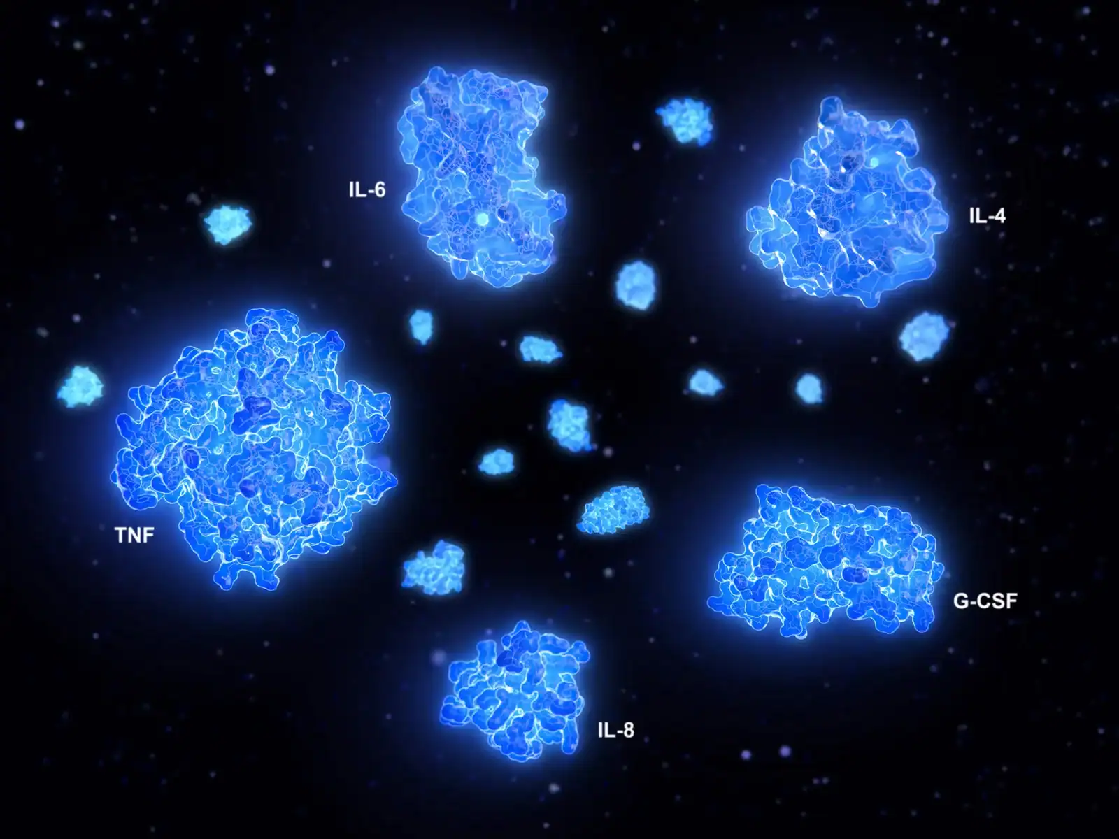
Because cytokines regulate communication between immune cells and tissues, cytokine profiling has applications across diseases involving altered immune activation, immune regulation, tissue responses or immune-cell trafficking.
Multiplex cytokine profiling can be used to investigate several dimensions of immune biology, including innate immune activation, T-cell differentiation, B-cell activity, interferon signaling, regulatory responses and immune-cell recruitment. In autoimmune disease research, these measurements can help researchers characterize disease-associated immune states and investigate how those states change over time or in response to treatment.
Importantly, cytokines should not generally be interpreted as isolated indicators of disease. Many are pleiotropic, meaning that they can act on multiple cell types and produce different effects depending on the cellular and tissue context. IL-2, for example, supports T-cell responses but is also critical for regulatory T-cell survival and immune tolerance. IL-10 is generally associated with immune regulation, yet its production can occur alongside inflammatory cytokines as part of the broader feedback response to immune activation. (3)
- In autoimmune disease research, this broader interpretation can be particularly valuable for investigating disease heterogeneity, identifying candidate immune signatures, characterizing patient subgroups and studying pharmacodynamic changes during treatment.
Why measure both immune-activating and regulatory cytokines?
Autoimmune disease is not simply a state of excessive immune activation. It can also involve impaired mechanisms that normally maintain self-tolerance and return immune responses toward homeostasis.
Regulatory T cells, regulatory B cells and immunoregulatory cytokines all contribute to controlling potentially damaging immune responses. Cytokines such as IL-2, IL-10 and TGF-β participate in these regulatory networks, although their effects depend on cellular context, concentration, timing and the surrounding cytokine environment. (4)
This creates an important consideration for cytokine profiling: an increase in cytokines involved in immune regulation should not automatically be interpreted as evidence that inflammation is resolving, just as an increase in an inflammatory cytokine does not by itself establish disease activity.

For example, IL-10 production can occur alongside inflammatory cytokine production as part of a feedback response intended to limit immune activation. Measuring IL-10 together with inflammatory and trafficking-related mediators may therefore provide additional information about the regulatory environment surrounding an immune response (3)
Similarly, IL-2 illustrates why cytokines cannot always be divided into simple “pro-inflammatory” and “anti-inflammatory” categories. IL-2 supports effector T-cell responses but is also essential for regulatory T-cell survival and function. In autoimmune disease research, measuring IL-2-related biology can therefore provide information about immune regulation and tolerance as well as immune activation. (1)
RA involves interconnected innate and adaptive immune pathways. TNF and IL-6 are important therapeutic and pathogenic pathways, but RA biology also involves Th1/Th17 responses, B-cell activity, chemokine-mediated recruitment and interactions between immune and stromal cells. Regulatory cytokines and mechanisms involved in resolution are also relevant to understanding why inflammation persists in some patients. (5, 6)
What is cytokine profiling?
Cytokine profiling is the simultaneous measurement of multiple cytokines, chemokines, and, depending on the assay, related growth factors or soluble immune mediators in biological samples.
Unlike single-analyte measurements, multiplex profiling enables researchers to examine several components of the immune response together. This is particularly useful because cytokines rarely function independently. Their effects depend on interactions between immune cells, receptor expression, local tissue conditions, and other signaling molecules.
For example, IL-6 and TNF contribute to inflammatory signaling in rheumatoid arthritis, while IL-17, IL-23, and other mediators influence adaptive immune responses and tissue inflammation. These pathways interact rather than operating as isolated mechanisms.
What can cytokine profiling reveal?
Three common applications in autoimmune disease research:
Immune pathway activity
Identify patterns associated with innate immunity, T-cell responses, interferon signaling, and immune regulation.
Longitudinal immune changes
Examine how circulating inflammatory mediators change with disease activity or treatment.
Biomarker discovery
Identify candidate proteins and combinations for subsequent validation.
Major cytokine pathways in autoimmune disease
Selecting cytokines for autoimmune disease research should begin with the biological pathway and research question rather than with the largest available cytokine panel.
Autoimmune diseases rarely depend on a single inflammatory mediator. Instead, cytokines operate in interconnected networks involving innate immune cells, T cells, B cells, stromal cells, and tissue-resident immune populations. The relative importance of these networks can differ not only between diseases, but also between patients with the same diagnosis, affected tissues, disease stages, and treatment exposures.
For this reason, cytokine profiling is generally most informative when analytes are interpreted as components of biological pathways, not as isolated biomarkers.
Cytokine pathways commonly investigated across autoimmune diseases
| Autoimmune disease / disease group | Dominant immune pathways investigated | Representative cytokines and mediators | Biological rationale for profiling |
|---|---|---|---|
| Rheumatoid arthritis (RA) | Innate immune activation, synovial tissue signaling, Th1/Th17 responses, B-cell activation, myeloid-cell recruitment | TNF-α, IL-6, IL-1β, IL-17A, IL-23, IFN-γ, GM-CSF, CXCL8, CCL2, BAFF | RA involves interactions between immune cells, synovial fibroblasts and inflammatory tissue environments. Cytokine profiling can help investigate inflammatory phenotypes, treatment response and biological heterogeneity between patients. TNF and IL-6 blockade demonstrate that cytokine pathways can be mechanistically important therapeutic targets. RA cytokine biology also includes Th17, chemokine and stromal pathways rather than a single inflammatory marker. |
| Systemic lupus erythematosus (SLE) | Type I interferon signaling, B-cell survival, autoantibody-associated immunity, immune tolerance failure | IFN-α, IFN-β, CXCL10, BAFF, APRIL, IL-6, IL-10, IL-17, TNF-α | SLE is one of the strongest examples of a systemic autoimmune disease where cytokine profiling can reveal immune-state differences. Interferon signatures, B-cell pathways and regulatory imbalance are frequently investigated. Type I interferon signatures are among the most reproducible molecular findings in SLE. BAFF and interferon pathways interact with B-cell survival and autoantibody-producing responses. |
| Psoriasis and psoriatic arthritis (PsA) | IL-23/Th17 differentiation, tissue-resident immune responses, keratinocyte/stromal signaling | IL-23, IL-17A, IL-17F, IL-22, TNF-α, IL-6, CXCL1, CXCL8 | Psoriasis demonstrates how cytokines regulate communication between immune cells and non-immune tissues. Cytokine profiling can investigate pathway activation and response to targeted therapies. IL-23/IL-17 biology is strongly supported by genetic studies and clinical responses to IL-17- and IL-23-targeted therapies. |
| Inflammatory bowel disease (Crohn’s disease and ulcerative colitis) | Mucosal immunity, epithelial barrier regulation, Th17/Th1 responses, innate immune activation | TNF-α, IL-23, IL-12, IL-17, IL-6, IL-1β, IL-10, TGF-β, CXCL8 | Cytokine profiling helps investigate intestinal immune regulation, inflammatory pathways and differences between disease phenotypes. Anti-TNF therapies demonstrate the importance of TNF signaling, while IL-12/23-targeted approaches demonstrate that multiple cytokine pathways contribute to intestinal immune dysregulation. |
| Multiple sclerosis (MS) | CNS immune-cell trafficking, Th1/Th17 responses, B-cell activity, neuroimmune communication | IFN-γ, IL-17A, IL-23, IL-6, GM-CSF, CXCL10, CCL2, BAFF | MS involves immune-cell migration across the blood–brain barrier and interactions between peripheral immune cells and CNS tissues. Th17-associated cytokines, chemokines and B-cell pathways are among the major immune mechanisms investigated in MS. |
| Sjögren’s syndrome | Type I interferon signaling, B-cell activation, glandular immune infiltration | IFN-α, CXCL10, BAFF, IL-6, IL-21, IL-10, TNF-α | Cytokine profiling may help investigate glandular immune activation, B-cell survival signals and systemic immune features. Sjögren’s syndrome shares interferon and BAFF-associated pathways with other systemic autoimmune diseases, particularly SLE. |
| Systemic sclerosis (scleroderma) | Fibrosis-associated signaling, immune activation, vascular responses, interferon pathways | IL-6, IFN-α, IFN-γ, TGF-β, CXCL10, IL-13, TNF-α | Unlike purely inflammatory diseases, systemic sclerosis requires assessment of immune signaling together with tissue remodeling and fibrosis pathways. Cytokines contribute to interactions between immune cells, fibroblasts and vascular tissues; IL-6 and interferon pathways are frequently investigated |
| Autoimmune thyroid diseases (Hashimoto thyroiditis, Graves’ disease) | Th1/Th17 responses, B-cell autoimmunity, antibody production | IFN-γ, IL-17A, IL-6, IL-10, BAFF, TGF-β | Cytokine analysis can help characterize immune polarization and B-cell-associated autoimmune responses. Studies identify altered Th1/Th17/regulatory cytokine balance in autoimmune thyroid conditions, although biomarkers require validation. |
| Autoimmune blistering diseases (pemphigus, pemphigoid) | Autoantibody production, B-cell activation, tissue immune injury | IL-4, IL-6, IL-17, TNF-α, IL-10, BAFF | Cytokine profiling may provide insight into antibody-producing immune responses and tissue inflammation. B-cell and cytokine pathways contribute to autoantibody generation and disease activity. |
| Autoimmune vasculitis (e.g., ANCA-associated vasculitis, giant cell arteritis) | Innate immune activation, neutrophils, vascular inflammation, T-cell responses | IL-6, IL-1β, TNF-α, IL-17, IFN-γ, CXCL chemokines | Cytokine profiles can help investigate vascular immune activation and inflammatory cell recruitment. IL-6, IL-1 and Th17-associated pathways are implicated in several vasculitic disorders. |
| Autoinflammatory diseases (CAPS, familial Mediterranean fever, Still’s disease) | Inflammasome activation, innate immune dysregulation | IL-1β, IL-18, IL-6, TNF-α, IL-1RA | These disorders demonstrate cytokine dysregulation driven primarily by innate immune mechanisms rather than classical autoantibody-mediated autoimmunity. IL-1 pathway activation is central in inflammasome-associated diseases, and IL-1-targeted therapies provide mechanistic validation. |
| Celiac disease and autoimmune gastrointestinal disorders | Barrier immunity, intraepithelial lymphocytes, Th1 responses | IFN-γ, IL-15, IL-21, IL-17, TNF-α | Cytokine profiling can investigate epithelial immune activation and adaptive immune responses to dietary antigens. IL-15 and T-cell activation pathways are central areas of investigation in celiac disease. |
| Idiopathic inflammatory myopathies (dermatomyositis, polymyositis, immune-mediated necrotizing myopathy) | Interferon signaling, muscle immune injury, adaptive immunity | IFNs, IFN-γ, IL-6, TNF-α, CXCL10 | Cytokines may reflect immune activation patterns associated with different disease subgroups. Interferon-associated pathways are particularly studied in dermatomyositis and related disorders. |
Why a pathway signature can outperform a single analyte?
There are several reasons a multi-analyte signature can be more informative than a single-marker result.
First, biological redundancy means the same downstream inflammatory phenotype can arise through multiple cytokines. Second, different disease subsets can reach similar clinical phenotypes through different upstream mechanisms. Third, circulating protein abundance is not identical to pathway activity: low serum IL-17A, for example, does not demonstrate absence of IL-17 activity within a joint, gut or skin lesion. Fourth, treatment itself changes the network, making a cytokine potentially more useful as a pharmacodynamic marker than as a diagnostic marker.
Why measure both immune-activating and regulatory cytokines?
Autoimmune disease is not simply a state of excessive immune activation. It can also involve impaired mechanisms that normally maintain self-tolerance and return immune responses toward homeostasis.
Regulatory T cells, regulatory B cells and immunoregulatory cytokines all contribute to controlling potentially damaging immune responses. Cytokines participate in these regulatory networks, although their effects depend on cellular context, concentration, timing and the surrounding cytokine environment.
This creates an important consideration for cytokine profiling: an increase in a regulatory cytokine should not automatically be interpreted as evidence that inflammation is resolving, just as an increase in an inflammatory cytokine does not by itself establish disease activity. (4)
Cytokine profiling solutions from Eve Technologies
For researchers investigating autoimmune disease, the choice of assay depends on whether the objective is broad immune profiling, targeted biomarker measurement, or confirmation of specific analytes. Eve Technologies offers multiplex cytokine and immune-profiling solutions designed to support these different stages of research.
For research applications, Eve Technologies provides multiplex cytokine assays that enable researchers to measure multiple immune mediators from a single biological sample. Broad panels can be used for exploratory studies of immune signaling, biomarker discovery and patient subgroup characterization, while more focused panels can support hypothesis-driven studies and longitudinal monitoring of selected pathways.
We also supports patient-focused testing through out CLIA-certified laboratory, offering high-dimensional immune profiling alongside its research assay services. The HD95 panel provides a broad assessment of cytokines and other immune-related biomarkers, enabling measurement of multiple immune signals from a patient sample rather than focusing on a single analyte. This high-dimensional approach can help characterize patterns of immune activity and regulation across a range of conditions. As with any laboratory test, results should be interpreted in the context of the specific test’s intended use, specimen type and analytical characteristics.
Conclusion
Cytokine profiling provides researchers with a way to investigate the interconnected immune pathways involved in autoimmune diseases. By measuring inflammatory, regulatory, and immune-cell recruitment signals together, researchers can explore disease-associated signatures that may be difficult to identify through individual biomarker measurements.
The most informative studies combine biologically justified biomarker selection with appropriate sample handling, fit-for-purpose assay performance, rigorous statistical analysis, and independent validation.
Whether investigating rheumatoid arthritis, systemic lupus erythematosus, psoriatic arthritis, or multiple sclerosis, the goal is to produce reproducible measurements that can support a more detailed understanding of disease biology.
For researchers planning an autoimmune disease study, Eve Technologies offers broad multiplex discovery panels and focused cytokine assays to support exploratory and targeted biomarker research.
Frequently asked questions
What is the difference between cytokine profiling and cytokine measurement?
Cytokine measurement can refer to quantifying one or several individual cytokines. Cytokine profiling generally involves measuring multiple immune mediators to investigate broader patterns of immune signaling.
Which cytokines are most important in autoimmune disease?
There is no universal cytokine panel for every autoimmune condition. Frequently investigated cytokines include TNF-α, IL-6, IL-1 family members, IL-17-family cytokines, interferons, IL-10, and selected chemokines, but their relevance depends on the disease, biological question and study design.
Can cytokine profiling distinguish autoimmune diseases?
Research studies have identified shared and disease-associated cytokine patterns. However, overlap between inflammatory pathways, patient heterogeneity, and analytical variability limit the use of exploratory profiles for distinguishing individual diseases. Such findings require independent validation before clinical application.
Is serum or plasma better for cytokine profiling?
The appropriate matrix depends on the analytes and assay. Plasma may reduce clotting-associated changes in some mediators, but neither matrix is optimal for every cytokine. Consistent sample collection and processing are essential.
Should researchers use a 96-plex panel or a smaller panel?
Broad panels are useful when the objective is exploratory biomarker discovery or the investigation of several immune pathways. Smaller panels may be more appropriate when the study has predefined targets or requires focused longitudinal measurements.
Can cytokine profiling measure treatment response?
Longitudinal cytokine profiling can help investigate changes in immune signaling during treatment. However, a change in cytokine concentration does not necessarily establish clinical response, therapeutic efficacy, or the mechanism responsible for an observed outcome.
What is cytokine profiling in autoimmune disease research?
Cytokine profiling is the measurement of multiple cytokines, chemokines and related immune mediators in a biological sample to identify pathway-level immune signaling patterns. In autoimmune research it is used for mechanism mapping, biomarker discovery, patient stratification and treatment-response monitoring. Its interpretation depends strongly on disease stage, tissue compartment, sample handling and assay platform.
Which platform is best for cytokine profiling?
There is no universally best platform. High-plex bead assays are efficient for broad discovery; ECL is attractive for focused, low-abundance panels; ELISA is useful for orthogonal validation; and mass spectrometry is best suited to enriched or targeted workflows when molecular specificity is more important than routine low-pg/mL sensitivity.
Should autoimmune cytokines be measured in serum or plasma?
The matrix should be chosen before the study and kept constant. Serum and plasma can yield substantially different values, particularly for platelet-associated mediators, so they should not be treated as interchangeable specimens.
Can a cytokine panel diagnose autoimmune disease?
Cytokine panels are powerful research tools, but most circulating cytokines are neither sufficiently disease-specific nor analytically standardized to serve as stand-alone diagnostic tests. Their strongest current role is pathway characterization, biomarker discovery, stratification and pharmacodynamic monitoring.
References:
- Hernandez, R., Põder, J., LaPorte, K.M. et al. Engineering IL-2 for immunotherapy of autoimmunity and cancer. Nat Rev Immunol 22, 614–628 (2022). https://doi.org/10.1038/s41577-022-00680-w
- O’Shea, J., Ma, A. & Lipsky, P. Cytokines and autoimmunity. Nat Rev Immunol 2, 37–45 (2002). https://doi.org/10.1038/nri702
- Saraiva, M., O’Garra, A. The regulation of IL-10 production by immune cells. Nat Rev Immunol 10, 170–181 (2010). https://doi.org/10.1038/nri2711
- Rubtsov, Y., Rudensky, A. TGFβ signalling in control of T-cell-mediated self-reactivity. Nat Rev Immunol 7, 443–453 (2007). https://doi.org/10.1038/nri2095
- McInnes, I., Buckley, C. & Isaacs, J. Cytokines in rheumatoid arthritis — shaping the immunological landscape. Nat Rev Rheumatol 12, 63–68 (2016). https://doi.org/10.1038/nrrheum.2015.171
- Chen, Z., Bozec, A., Ramming, A. et al. Anti-inflammatory and immune-regulatory cytokines in rheumatoid arthritis. Nat Rev Rheumatol 15, 9–17 (2019). https://doi.org/10.1038/s41584-018-0109-2
- Psarras, A., Wittmann, M. & Vital, E.M. Emerging concepts of type I interferons in SLE pathogenesis and therapy. Nat Rev Rheumatol 18, 575–590 (2022). https://doi.org/10.1038/s41584-022-00826-z
- Mihaescu G, Gradisteanu Pircalabioru G, Roznovan CN, Ditu LM, Comanici MM, Savu O. Interferons in Autoimmunity: From Loss of Tolerance to Chronic Inflammation. Biomedicines. 2025 Oct 11;13(10):2472. doi: 10.3390/biomedicines13102472.
- Majumder S, McGeachy MJ. IL-17 in the Pathogenesis of Disease: Good Intentions Gone Awry. Annu Rev Immunol. 2021;39:537-556. doi: 10.1146/annurev-immunol-101819-092536.