- Discovery Assays

Human

Mouse
Rat
Porcine
Canine
Equine
Sheep
Bovine

Chicken
- Custom Plex Assays

Human
Mouse
Rat
Porcine
Canine
Equine

Sheep

Bovine
Chicken
- My Account
-


Multiplex Cytokine Profiling in Cancer Immunotherapy: Uncovering Tumor Cytokine Biomarkers and Monitoring Cytokine Release Syndrome
Introduction
Cancer immunotherapies harness the patient’s immune system, but individual responses vary widely. Measuring cytokines – small immune‐signaling proteins – can reveal how a patient’s immune system is reacting to therapy.
Measuring cytokines such as interleukin‑6 (IL‑6) and interferon‑γ (IFN‑γ) reveals critical biomarkers involved in anti‑tumor activity, while their dysregulated release can cause life‑threatening cytokine release syndrome (CRS), also known as a “cytokine storm.” For more on cytokine storm, read Cytokine Release Syndrome (CRS) Explained: Key Insights for Clinicians.
Multiplex cytokine profiling offers a high-throughput, comprehensive snapshot of the tumor microenvironment and systemic immune activation by measuring dozens of cytokines simultaneously from a single small sample (e.g., Luminex® panels). These systems use color-coded beads bound to unique antibodies to detect and quantify multiple cytokines with high sensitivity and precision.
Eve Technologies offers multiplex assays commonly used in immunotherapy research, allowing simultaneous analysis of IL‑6, IFN‑γ, and other key cytokines. You can view our full list of human cytokine assays and our CLIA-certified clinical tests.
In this post we will explore why cytokine profiling in cancer immunotherapy matters, highlight key biomarkers driving outcomes, and outline how multiplex cytokine assays can accelerate your research and clinical insights.
Why Multiplex Cytokine Profiling Matters in Cancer Immunotherapy
Holistic Immune Snapshot
Traditional assays like ELISA measure one cytokine at a time, using large sample volumes. These remain popular as most labs have the labware and the technical expertise to run these assays in house, so they’re good for initial discovery. In contrast, bead‑based multiplex platforms can profile dozens of cytokines in parallel from as little as 25 µL of serum or plasma. This lets researchers map the full cytokine network (“immune fingerprint”) in patients. Larger plexes generate larger data sets, which also lend themselves well to multi-variate machine learning data analysis (e.g., PCA). Multiplex systems like Luminex® use color-coded beads, each bound to a unique antibody, to detect and quantify multiple cytokines simultaneously with high sensitivity and precision.
To learn more about which platform to choose for multiplex cytokine profiling, read our blog post Multiplex Cytokine Assays Compared: Luminex®, Meso Scale Discovery®, and Traditional ELISA.
Time and Cost Efficiency
Multiplex panels drastically reduce both labor and expense. By measuring dozens of cytokines in a single run, they eliminate the need for multiple plates, repeated pipetting, and excess sample volume. Compared to singleplex assays like ELISA, multiplex immunoassays deliver greater throughput, higher efficiency, and lower assay cost per analyte. In fact, studies show they can reduce overall assay time by over 80% and cost per marker by a similar margin. This makes large-scale immune profiling feasible, even under tight timelines and budgets.
The comparison below illustrates the difference. To measure 48 analytes using traditional ELISA, you would need 48 separate 96-well plates and over 100 hours of labor. In contrast, a multiplex assay requires just one plate and a few hours of work, while delivering greater sensitivity and high-quality data in the picogram range.

- That’s why we offer our Discovery Assays: comprehensive, Luminex®-based panels designed for immune profiling in cancer, inflammation, and more. They’re also highly affordable thanks to our pay-per-well pricing model, so you only order what you need.
Whether you’re studying 15 markers, 48, or even 96, multiplexing offers unmatched scalability. It’s ideal for researchers needing both depth and efficiency in immune monitoring and biomarker discovery.

Improved Sensitivity and Dynamic Range
Modern multiplex platforms (especially high-sensitivity Luminex® or electrochemiluminescence systems like the MSD®) detect cytokines at pg/mL levels, capturing even weak signals. For example, one multisite comparison showed that high-sensitivity panels could reveal cytokine fluctuations “not previously recognized” (1). Detecting low-abundance cytokines early is crucial for recognizing subtle immune activation or suppression in patients. Multiplex kits typically span a broad dynamic range, allowing both low- and high-abundance biomarkers to be quantified in the same assay.
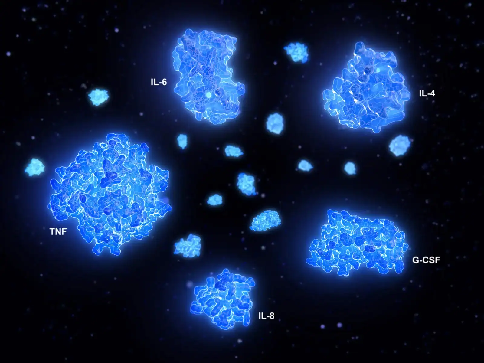
Key Cytokines Driving Immunotherapy Outcomes
Understanding which cytokines to monitor is the first step in effective cytokine profiling immunotherapy:
| Cytokine | Role | Clinical Insight |
| IL‑6 | Pro‑inflammatory mediator | Elevated IL‑6 is the hallmark of severe cytokine release syndrome (CRS) in CAR‑T therapy and correlates with toxicity in checkpoint inhibitor treatments. Early IL‑6 spikes can trigger timely interventions (e.g., tocilizumab). |
| IFN‑γ | Th1 activation | High IFN‑γ levels indicate robust anti‑tumor T‑cell responses. Tracking IFN‑γ alongside IL‑6 helps distinguish effective immune activation from harmful inflammation. |
| IL‑10 | Regulatory cytokine | Paradoxically, elevated IL‑10 can predict immune‑related adverse events (irAEs) such as pneumonitis in ICI‑treated lung cancer patients. Monitoring IL‑10 enables early risk stratification. |
| IL‑2 | T‑cell proliferation | Pre‑treatment IL‑2 levels have been linked to better checkpoint inhibitor outcomes, making IL‑2 a valuable immunotherapy response biomarker. |
| TNFα | Acute inflammation | Early TNFα rises often coincide with CRS onset. Multiplex panels that include TNFα and IL‑6 are essential for comprehensive cytokine release syndrome monitoring. |
Spotlight on CAR‑T Therapy and Checkpoint Inhibitors
CAR‑T Cell Therapy
CAR‑T treatments (e.g. CD19 CAR‑T for ALL or DLBCL, BCMA CAR‑T for myeloma) have revolutionized blood cancer care. However, about 30–50% of patients experience CRS (any grade) (2), which can be life‑threatening if unchecked. Real‑time cytokine monitoring with a broad panel greatly improves safety and informs personalized intervention:
- Early Detection of CRS: Cytokines rise hours to days before clinical symptoms. For example, IL‑6 often climbs 0–36 hours post‑infusion, even before fever appears (2). Catching this early rise allows preemptive action.
- Personalized Intervention: Guiding the use of IL‑6 blockers or corticosteroids to prevent escalation.
- Outcome Correlation: Linking cytokine trajectories with therapeutic efficacy and long‑term remission rates.
For research use, we offer our HDF15 and HD96 Discovery Assays, with HD96 being one of the most comprehensive cytokine panels available and HDF15 providing a focused immune profile. Both are also available as CLIA-certified tests for patient diagnostics (HDF15-Clin and HD95-Clin) and support early detection of CRS.
Checkpoint Inhibitors (ICIs)
Immune checkpoint inhibitors (anti‑PD1/PDL1, anti‑CTLA4) frontline treatments in melanoma, NSCLC, and other solid tumors. Multiplex profiling aids their use by identifying biomarkers of response and toxicity:
- Predictive Biomarkers: Studies have linked dynamic cytokine changes to ICI success. For instance, a 2025 meta‑analysis found that high IL‑6 and IL‑10 at baseline were associated with shorter progression‑free survival in NSCLC patients on PD‑1 therapy (3). Conversely, a strong IFN‑γ signature in the tumor microenvironment predicts response. In another NSCLC cohort, high baseline IL‑2 (HR=0.67) and high soluble PD‑L1 (sPD‑L1) markedly predicted longer PFS, while IL‑23 tracked with poorer outcome (4). Overall, multiplex panels can pick up early signs – e.g. an IL‑6 spike or IL-10 rise – that forecast benefit or resistance.
- Toxicity Management: As noted above, IL‑10 is a warning sign for irAEs. Patients with rising IL‑10 during therapy often go on to develop pneumonitis or colitis (5). Monitoring cytokines lets oncologists stratify risk: those with adverse cytokine profiles can be monitored more intensively or receive lower immunotherapy dosing.
- Therapeutic Adjustment: Cytokine profiles also inform combination strategies. For example, a high Th1/Th2 imbalance (e.g. rising IFN‑γ with low IL‑4/IL‑10) suggests a hot tumor that may need only ICI, whereas a strongly immunosuppressive profile (high IL-10, TGF-β) might justify adding cytokine modulators or vaccines. By mapping Th1 (IFN‑γ, IL‑2) vs. regulatory (IL-10, TGF-β) signatures, researchers tailor the next steps – such as adding an IL-2 agonist or blocking IL-10 pathways.
Identifying Tumor Cytokine Biomarkers
Beyond blood, tumors themselves secrete cytokines that reflect the tumor microenvironment. Multiplex panels can help discover such tumor cytokine biomarkers:
- Angiogenic Markers: Cytokines like VEGF, FGF, and angiopoietins signal blood vessel growth. High VEGF levels often correlate with advanced cancer stage and metastasis (6). For example, meta-analyses show VEGF expression is significantly higher in tumors that are high grade or stage (6). Tracking these in patient samples can indicate tumor vascular activity and aggressiveness.
- Immunosuppressive Factors: TGF‑β and IL‑10 often indicate an immunosuppressive (“cold”) niche. Elevated TGF‑β in the tumor microenvironment is linked to poor ICI response, while IL‑10 is produced by Tregs and M2 macrophages. Measuring these helps identify tumors that might resist immunotherapy.
- Inflammatory Signatures: Cytokines like IL‑17 or IL‑23 mark active inflammation. For instance, IL‑17 pathways have been tied to checkpoint inhibitor pneumonitis. Patients with high tumor IL‑17 or IL‑23 may benefit more from PD‑1 inhibitors but also risk specific toxicities.
How Eve Technologies Supports Your Research
Eve Technologies offers flexible multiplex cytokine services to fit any study:
Predefined Discovery Assays
Discovery Assays are preconfigured multiplex panels with a set number of analytes, available through our flexible pay-per-well pricing model. We offer panels ranging from 3-plex up to 96-plex to suit different research needs. Choose the assay that fits your budget and research goals without paying for unused wells.
Custom Plex Options
Build your own panel with any combination of analytes. Add or remove targets based on your hypothesis, no minimum analyte count is required. This lets you tailor an assay exactly to your research needs (e.g., include novel tumor markers or exclude irrelevant ones).
Flexible Sample Types
Run assays on serum, plasma, cell culture supernatant, or even tissue lysate with minimal prep.
Fast Turnaround Time
Industry-leading processing times mean you get results quickly, keeping your project on schedule.
Whether you’re scouting new immunotherapy biomarkers, optimizing CAR‑T protocols, or profiling the tumor microenvironment, Eve Technologies’ multiplex cytokine panels give you the breadth and depth needed to uncover actionable insights.
Sit back, relax, and focus on what matters most—your research. While you dive into discovery, we’ll handle the assays with precision, consistency, and care. Let us do the benchwork so you can do the breakthrough.
Take the Next Step: Drive Discoveries with Multiplex Cytokine Profiling
Whether you’re investigating novel immunotherapy response biomarkers, optimizing CAR‑T protocols, or mapping tumor cytokine biomarkers, Eve Technologies’ multiplex cytokine panels deliver the depth and flexibility you need:
- Get a Quote: Visit Eve Technologies’ Discovery Assays page to explore predefined panels or start designing a custom array tailored to your hypothesis.
- Submit Your Samples: Mail your serum, plasma, or other samples to us using our simple submission process.
- Accelerate Insights: Leverage our expert team and robust analytics to turn complex cytokine data into clear biological conclusions and translational discoveries.
Unlock the full potential of cytokine profiling in cancer immunotherapy – contact Eve Technologies today and bring precision immune biomarker analysis to your next oncology study.
Discover more blog posts:
- CLIA-Certified Cytokine Testing for Physicians and Referring Labs
- Cytokine Testing for Clinical Trials: Multiplex Biomarker Support for Early Phase Studies
- Outsourcing Cytokine Testing vs In-House: Cost, Time, and Data Quality Compared
- Custom Multiplex Cytokine Panels: What Researchers Should Consider
- Ultrasensitive Immunoassays Breakthrough: Detecting Low-Abundance Biomarkers
References and Resources
- Breen, E. C., Reynolds, S. M., Cox, C., Jacobson, L. P., Magpantay, L., Mulder, C. B., Dibben, O., Margolick, J. B., Bream, J. H., Sambrano, E., Martínez-Maza, O., Sinclair, E., Borrow, P., Landay, A. L., Rinaldo, C. R., Norris, P. J., … Norris, P. J. (2011). Multisite comparison of high-sensitivity multiplex cytokine assays. Clinical and Vaccine Immunology, 18(8), 1229–1242. https://doi.org/10.1128/CVI.05032-11
- DePriest, B. P., Vieira, N., Bidgoli, A., & Paczesny, S. (2021). An overview of multiplexed analyses of CAR T-cell therapies: Insights and potential. Expert Review of Proteomics, 18(9), 767–780. https://doi.org/10.1080/14789450.2021.1992276
- Liu, Q., Shaibu, Z., Xu, A., Yang, F., Cao, R., & Yang, F. (2025). Predictive value of serum cytokines in patients with non-small-cell lung cancer receiving anti-PD-1 blockade therapy: A meta-analysis. Clinical and Experimental Medicine, 25(1), 59. https://doi.org/10.1007/s10238-025-01587-1
- Jain, K., Goel, A., Mehra, D., Rathore, D. K., Binayke, A., Aggarwal, S., Ganguly, S., Awasthi, A., Madan, E., & Ganguly, N. K. (2025). Cytokine profiling identifies circulating IL-2, IL-23 and sPD-L1 as prognostic biomarkers for treatment outcomes in non-small cell lung cancer patients undergoing anti-PD1 therapy. Frontiers in Oncology, 15, Article 1628379. https://doi.org/10.3389/fonc.2025.1628379
- Wang, H., Zhou, F., Zhao, C., Cheng, L., Zhou, C., Qiao, M., Li, X., & Chen, X. (2022). Interleukin-10 is a promising marker for immune-related adverse events in patients with non-small cell lung cancer receiving immunotherapy. Frontiers in Immunology, 13, Article 840313. https://doi.org/10.3389/fimmu.2022.840313
- Cai, C., Wang, X., Fu, Q., & Chen, A. (2022). The VEGF expression associated with prognosis in patients with intrahepatic cholangiocarcinoma: A systematic review and meta-analysis. World Journal of Surgical Oncology, 20, Article 40. https://doi.org/10.1186/s12957-022-02511-7